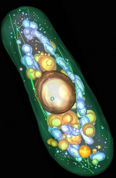
Scientists produce the first high resolution 3D image of a complete eukaryotic cell
Researchers at the European Molecular Biology Laboratory (EMBL) and the University of Colorado have now obtained the first 3-D visualization of a complete eukaryotic cell at a resolution high enough to resolve the cytoskeleton’s precise architectural plan in fission yeast.
The electron tomogram of a complete yeast cell reveals the cellular architecture. It shows plasma membrane, microtubules and light vacuoles (green), nucleus, dark vacuoles and dark vesicles (gold), mitochondria and large dark vesicles (blue) and light vesicles (pink).
References:
- Hoog JL, Schwartz C, Noon AT, O’toole ET, Mastronarde DN, McIntosh JR, Antony C. Organization of interphase microtubules in fission yeast analyzed by electron tomography. Dev Cell. 2007 Mar;12(3):349-61. Pubmed


[…] A nice picture can be found at Migrations […]
By: YourSciCom » 3D-Reconstruction of a Complete Eukaryotic Cell on March 7, 2007
at 11:22 am
[…] tomography has also been used to construct a three-dimensional visualization of a complete eurkarotic cell (S. pombe). The power of such high-power resolution to confirm sequence homologies with functional […]
By: Seeing Bacterial Bones with Cryo-EM Tomography | Bitesize Bio on January 14, 2008
at 7:32 am
Wow.
By: Brooke on December 7, 2009
at 4:52 am